

I agree Our site saves small pieces of text information (cookies) on your device in order to deliver better content and for statistical purposes. You can disable the usage of cookies by changing the settings of your browser. By browsing our website without changing the browser settings you grant us permission to store that information on your device.
See our Anatomopathology dedicated page to explore all our available tests.
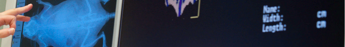
Gait analysis evaluates proprioceptive sensitivity but also assesses the integrity of motor coordination and vestibular function. Animals require an accurate paw placement to succeed in this test. The gait is altered in in different pathologies including ataxia or neuropathic pain.
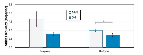
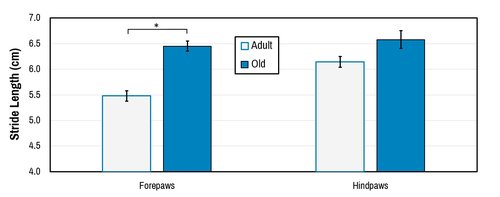
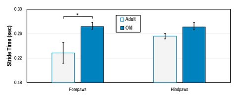
Sample test results:
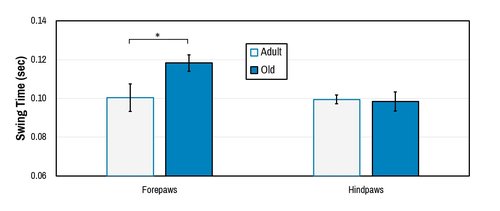
Hansen and Pulst, 2013.
Digigait™ (Mouse Specifics, Inc.) transparent treadmill instrumentation.
10 mice per group are recommended for reliabe data analysis.
The balance beam test or gait analysis is relatively specific for evaluation of proprioceptive sensitivity. However, the performance in this test also depends on the integrity of motor coordination and vestibular function.
An elevated beam of standardized diameter and length.
10 mice per group are recommended for reliable data analysis.
Electromyographic recording allows neurophysiological measurement of sensory-motor function. In situations of demyelination and/or axonal degeneration, the compound muscle action potential (CMAP), latency of motor response and sensitive nerve conduction velocity (SNCV) are affected.
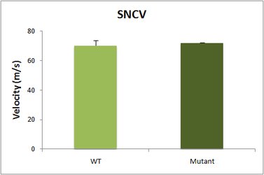
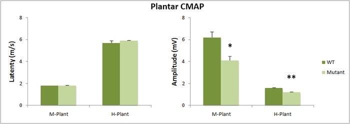
Key Point electromyograph (EMG) apparatus (Medtronic, Boulogne-Billancourt, France). The body temperature is maintained at 37 °C with a homeostatic blanket (Harvard, LES ULIS, France).
10 mice per group are recommended for reliable data analysis.
This test measures the muscular muscle strength using an isometric dynamometer connected to a grid. Once the animal is holding the grid with its forepaws it is slowly moved backwards until it releases it. The dynamometer records the maximal force exerted.
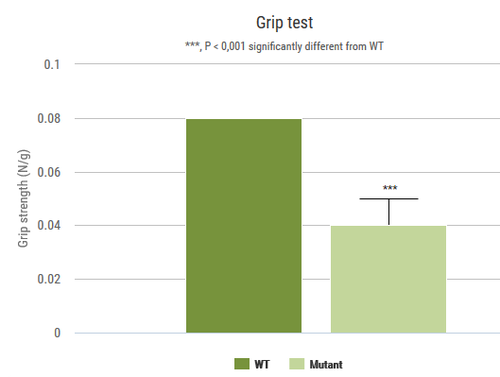
2 grip strength apparati (BIOSEB, Vitrolles, France).
10 mice per group are recommended for reliable data analysis.
Muscle contractility and muscle force can be evaluated in situ on limb muscle using an isolated muscle test system (Aurora Scientific). This procedure is done on anesthetized mice, animals are sacrificed at the end of the measurement.
A proprioceptive test similar to the balance beam under more challenging condition requiring vertical as well as lateral movement. This test also depends on the integrity of motor coordination and vestibular function.
The one-meter swim test is used to evaluate swimming ability of mice.
A straight swim tank (100 cm long, 6 cm wide, 27.3 cm high) made of translucent PVC and filled at 21 cm depth with water. A visible platform is placed opposite to the starting point, allowing to the animal to escape from water.
10 mice per group are recommended for reliable data analysis.
This test measures the ability of an animal to maintain balance on a rotating rod. This task requires a variety of proprioceptive, vestibular and fine-tuned motor abilities.
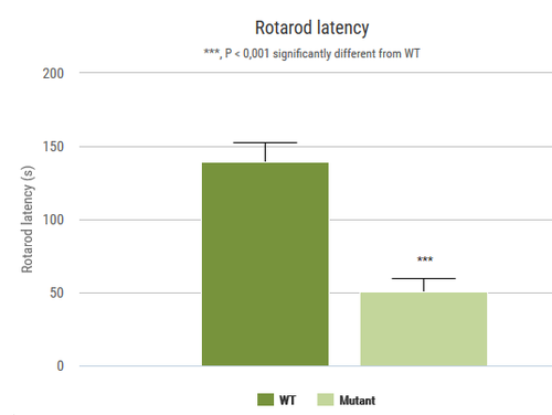
2 rotarod apparati (BIOSEB, Vitrolles, France)
10 mice per group are recommended for reliable data analysis.
The string test measures the time required for a forelimb-hanging mouse to gain hindlimb traction. Defects in latency time reflect potential alteration in traction force, drug-induced sedation, or deficient coordination (Meziane et al. 1996).

Customized equipment: a wire stretched horizontally 40 cm above a table.
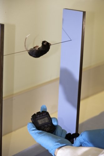
10 mice per group are recommended for reliable data analysis.
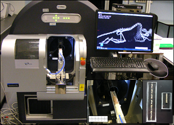
Bone architecture may be analysed by both in vivo and in situ using Micron-scale X-ray Computed Tomography (also called µCT). It is particularly helpful for bone dynamics analysis. This augments our current capacities of skeletal examinations by TRAP histological stain, X-ray, DEXA scanning, quantitative NMR, and adapted clinical chemistry analysis.
µCT (PerkinElmer Quantum FX, Waltham, Massachusetts)
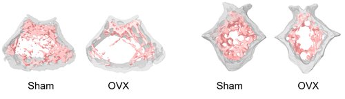
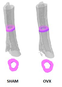
Representative 3D volumes illustrating trabecular microarchitecture in the distal femur (left) and caudal vertebra (right) of Sham-operated and Ovarectomized (OVX) mice, 9 weeks after surgery. Similar volumes are used for morphometric quantification.
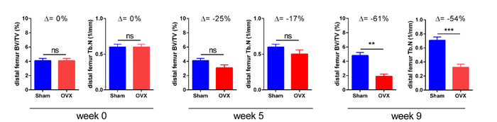
Example of longitudinal changes in trabecular bone volume fraction (BV/TV) and trabecular number (Tb.N) for Sham and OVX groups.
Representative 3D volumes illustrating cortical bone in the tibia of Sham-operated and Ovarectomized OVX mice, 9 weeks after surgery. The illustrations were generated by Alexandru Parlog (PHENOMIN-ICS).
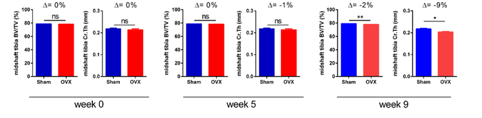
Example of longitudinal changes in cortical bone volume fraction (BV/TV) and cortical thickness (Cr.Th) for Sham-operated and Ovarectomized OVX groups.
Contact us for help in designing your experiments on bone dynamics.
The X-Ray system gives very precise images of the skeletton. In DXA mode it automatically calculates BMD, BMC, and lean and fat mass percentages.
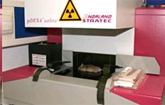
pDEXA Sabre (Norland)

CT is a technique that relies on differential levels of X-ray attenuation by tissues within the body to produce images reflecting anatomy.
The assay is developed to provide high resolution images of the whole mouse skeleton. Analysis of the digital X-ray pictures is generally performed with respect to bones from the head (zygomatic bone, maxilla, mandibles), teeth, scapulae, clavicle, ribs (number, shape, fusion), pelvis, vertebrae (numbers, shape and potential fusion of cervical, thoracic, lumbar, pelvic and caudal ones), limb bones (humerus, radius, ulna, femur, tibia), joints, digits and syndactylism.
X-Ray MX-20 Specimen (Faxitron, Tucson, Arizona, USA)
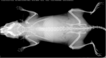
At least 5 mice per group are required for appropriated analysis.
Body composition for fat, lean and free body fluid is evaluated on conscious mice by quantitative nuclear magnetic resonance on Minispec analyzer.
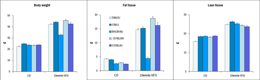
Body composition analysis of male mice from different common laboratory strains after provision of chow diet (CD) or 10 weeks provision of high-fat diet (HFD), including complete body weight, fat tissue content, and lean tissue content. Dramatic increases in fat tissue is seen upon HFD-diet treatment for most strains excepting BALB/c, consistent with what has previously been reported.
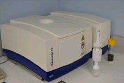
Body composition is evaluated by Quantitative Nuclear Magnetic Resonance (qNMR) using Minispec+ analyzer (Bruker BioSpin S.A.S., Wissembourg, France)
See our Gene expression analysis dedicated page to explore our different related tests.
The High-Resolution Digital Radiography system (Ultrafocus by Faxitron) gives very precise images of the skeletton. Analysis of the digital X-ray pictures is generally performed with respect to bones from the head (zygomatic bone, maxilla, mandibles), teeth, scapulae, clavicle, ribs (number, shape, fusion), pelvis, vertebrae (numbers, shape and potential fusion of cervical, thoracic, lumbar, pelvic and caudal ones), limb bones (humerus, radius, ulna, femur, tibia), joints, digits and syndactylism.
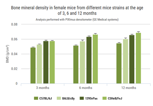
In dual-energy x-ray absorptiometry mode (DXA), the system measures bone mineral density (BMD) and also determine lean and fat mass percentages.

Comparison of wild-type C57BL/6N (WT) and Ttc39a deletion (Mutant) mice for noted body composition parameters by non-invasive Ultrafocus DXA imaging. *, p<0.05; **, p<0.01.
See our In vivo Viral Transduction dedicated page to explore our frequently used vectors and delivery modes including stereotactic admininistration (intraventricular and cerebellar)
With several months notice, we can procure and provide a variety of different restriction diets for nutritional analyses in conjuction with our other analyses.
With several months notice, we can procure and provide a variety of different restriction diets for nutritional analyses in conjuction with our other analyses.
See our In vivo Viral Transduction dedicated page to explore our frequently used vectors and delivery modes including stereotactic admininistration (intraventricular and cerebellar)